

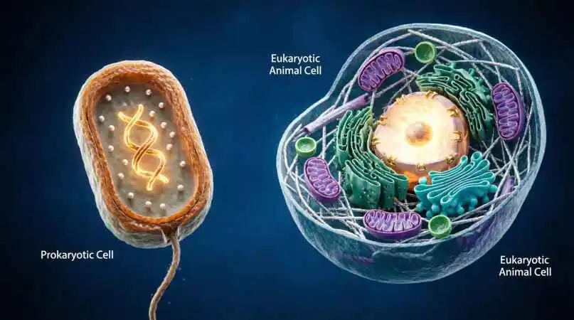
The Ultimate Guide to Eukaryotic and Prokaryotic Cells — The Microscopic Forces That Built All Life on Earth
Every living thing that has ever existed — from the first bacterial speck in a primordial ocean 3.8 billion years ago to the 37 trillion cells humming inside your body right now — is built from one of two fundamental cell designs. Understanding these two blueprints is not just biology class material. It is the story of how life itself was invented, diversified, and conquered an entire planet.
Introduction: The Two Great Kingdoms of the Cellular World
Imagine you are an architect presented with every building ever constructed on Earth and asked to classify them. You study thousands of structures — huts, skyscrapers, temples, factories — and eventually you realize: despite infinite variation in size, material, and purpose, every single building falls into one of two categories. Either it has an organized central core that controls everything, or it is a single open room where everything coexists without separation.
This is precisely what scientists discovered when they looked at cells under increasingly powerful microscopes. Despite the staggering diversity of life — bacteria and blue whales, mushrooms and maple trees, amoebas and astronauts — every cell belongs to one of two fundamental types: prokaryotic (without a true nucleus) or eukaryotic (with a true nucleus).
This distinction is arguably the most important dividing line in all of biology. It separates organisms that arose nearly 4 billion years ago from those that appeared roughly 1.5 to 2 billion years later. It divides the simple from the complex, the ancient from the evolved, the invisible from the visible. And understanding it unlocks the logic of all life.
Part I: Prokaryotic Cells — Life’s Original Blueprint
What “Prokaryote” Actually Means
The word prokaryote comes from the Greek pro (before) and karyon (nucleus). These are cells that existed before the nucleus was invented by evolution — and they continue to exist today in almost incomprehensible numbers. Every bacterium and every archaeon on Earth is a prokaryote.
To appreciate just how ancient and successful prokaryotes are, consider this: they colonized Earth approximately 3.8 billion years ago when the planet was still geologically violent, largely anaerobic, and bombarded by ultraviolet radiation. Not only did they survive — they thrived, and they transformed Earth’s atmosphere, oceans, and chemistry in ways that made all subsequent life possible.
The Architecture of Simplicity
Do not mistake simplicity for weakness. A prokaryotic cell is an engineering masterpiece of minimalism — every component precisely calibrated for maximum efficiency in a minimal space.
Cell Size: Prokaryotic cells are typically 1–10 micrometers in diameter. To put this in perspective, you could line up approximately 1,000 average bacteria across the width of a single human hair.
The Cell Wall: Almost all prokaryotes have a cell wall surrounding the plasma membrane. In bacteria, this wall is made of peptidoglycan — a mesh-like polymer unique to bacteria that gives the cell its shape, prevents osmotic bursting, and is, crucially, the target of many antibiotics including penicillin. Archaea have cell walls made of different materials, further distinguishing them from bacteria.
No Membrane-Bound Organelles: This is the defining architectural feature of prokaryotes. There is no nucleus, no mitochondria, no endoplasmic reticulum, no Golgi apparatus — none of the membrane-enclosed compartments that define eukaryotic complexity. The entire cell is one fluid-filled compartment called the cytoplasm, in which all cellular processes occur simultaneously.
The Nucleoid Region: Though prokaryotes lack a true nucleus, they are not without genetic organization. Their DNA — typically a single, circular chromosome — is concentrated in a region called the nucleoid. This DNA is not enclosed by a membrane; it is simply coiled and compacted in a specific area of the cytoplasm. Additionally, many bacteria carry small circular DNA molecules called plasmids, which often carry genes for antibiotic resistance and can be transferred between cells.
Ribosomes: Prokaryotes have ribosomes — the molecular machines that build proteins — but their ribosomes are smaller (70S) than eukaryotic ribosomes (80S). This size difference is medically significant: many antibiotics (streptomycin, erythromycin, tetracycline) specifically target prokaryotic 70S ribosomes, killing bacteria without harming the host’s eukaryotic cells.
Flagella and Pili: Many prokaryotes are mobile, propelled by flagella — whip-like protein structures that rotate like a molecular motor. Pili are shorter, hair-like projections used for attachment to surfaces and, in some bacteria, for the transfer of DNA between cells (conjugation).
Plasma Membrane: The inner boundary of the prokaryotic cell is the plasma membrane — a phospholipid bilayer studded with proteins that regulates what enters and exits the cell, performs cellular respiration (since there are no mitochondria), and hosts many enzymatic reactions.
The Two Domains of Prokaryotes: Bacteria and Archaea
For most of the history of biology, “prokaryote” was treated as a single category. Then in the 1970s, microbiologist Carl Woese used ribosomal RNA sequencing to reveal a stunning truth: prokaryotes are actually two completely distinct domains of life, more different from each other than either is from eukaryotes in certain molecular ways.
Bacteria are the prokaryotes most familiar to us — E. coli in our guts, Streptococcus in our throats, Mycobacterium tuberculosis in lungs, Lactobacillus in yogurt, Rhizobium in soil fixing nitrogen. They are the most numerous organisms on Earth, with estimates suggesting 10^30 bacterial cells in the world’s oceans alone.
Archaea are the great survivors — often found in extreme environments once thought incompatible with life. Thermophiles thrive in volcanic hot springs at temperatures above 80°C. Halophiles colonize salt lakes where nothing else survives. Methanogens live in oxygen-free environments, producing methane as a metabolic byproduct. But archaea are not confined to extremes: they are abundant in normal soils, ocean water, and in the human digestive system.
Reproduction: The Prokaryotic Speed Advantage
Prokaryotes reproduce primarily through binary fission — a simple process in which the cell replicates its DNA and divides into two identical daughter cells. In optimal conditions, E. coli can complete this process in as little as 20 minutes. This means a single bacterium could theoretically become over 4 billion bacteria in 10 hours — explaining how bacterial infections can escalate rapidly and why bacterial evolution (and antibiotic resistance) operates on such compressed timescales compared to eukaryotic evolution.
Why Prokaryotes Rule the Earth
Despite their microscopic scale and structural simplicity, prokaryotes are unquestionably the dominant life form on Earth by every metric: total number, total biomass, metabolic diversity, and range of habitats. They were here first. They will almost certainly be here last. They drive the nitrogen cycle, the carbon cycle, the sulfur cycle — the fundamental chemical processes that keep Earth habitable for all other life. Our guts contain trillions of bacteria without which we could not digest food, synthesize vitamins, or train our immune systems. Prokaryotes are not the background of life’s story. They are the story.
Part II: Eukaryotic Cells — The Architecture of Complexity
The Great Leap: The Origin of the Eukaryote
Approximately 1.5 to 2 billion years ago, something happened in the history of life that changed everything: a prokaryotic cell acquired a nucleus. The precise mechanism is debated, but the result was the first eukaryote — a cell of dramatically greater size, complexity, and versatility than anything that had come before.
The word eukaryote comes from the Greek eu (true) and karyon (nucleus). These are cells with a true, membrane-enclosed nucleus — a protected inner sanctum where the cell’s genetic blueprint is stored, protected, and transcribed. Every plant, animal, fungus, and protist on Earth is made of eukaryotic cells.
The most widely accepted explanation for eukaryotic origins is the endosymbiotic theory, championed by biologist Lynn Margulis in the 1960s. According to this theory, the ancestors of mitochondria and chloroplasts were once free-living prokaryotes that were engulfed by a larger cell and, rather than being digested, established a permanent mutualistic relationship. The evidence for this is overwhelming: mitochondria and chloroplasts have their own DNA, their own ribosomes (of the prokaryotic 70S type), and reproduce by binary fission — all signatures of their ancient bacterial ancestry.
The Nucleus: Command and Control
The defining feature of the eukaryotic cell is the nucleus — a membrane-enclosed compartment that houses the cell’s DNA. This is not a minor organizational convenience. It is a profound biological innovation.
The nuclear envelope is a double membrane perforated by thousands of nuclear pores — complex protein structures that act as selective gates, allowing specific molecules (messenger RNA, proteins) to pass in and out while maintaining the nucleus as a protected environment.
Inside the nucleus, DNA is not naked. It is wrapped around proteins called histones, compacted into structures called nucleosomes, and organized into chromatin — a dynamic packaging system that regulates gene expression by controlling which sections of DNA are physically accessible for transcription.
Within the nucleus is the nucleolus — a dense region where ribosomal RNA is synthesized and ribosome assembly begins. Cells with high protein production needs (liver cells, secretory cells) have particularly large, prominent nucleoli.
The separation of DNA from the rest of the cell — the separation of transcription from translation — is far more than organizational housekeeping. It creates a regulatory delay that allows cells to quality-check RNA before it is translated into protein, enabling more sophisticated gene regulation than is possible in prokaryotes.
The Organelle System: A City of Specialists
What makes the eukaryotic cell so powerful is its division of labor. Rather than cramming all cellular functions into one undifferentiated space, the eukaryotic cell partitions activities into specialized membrane-enclosed compartments — organelles. Each organelle maintains a distinct internal chemistry optimized for its function.
Mitochondria are the cell’s power plants. Through the process of cellular respiration — oxidative phosphorylation — they generate ATP (adenosine triphosphate), the universal currency of cellular energy. A typical human cell contains hundreds to thousands of mitochondria, and highly active cells (heart muscle, liver cells) contain more than quieter cells. Their inner membrane is elaborately folded into cristae, maximizing the surface area available for ATP synthesis. As noted, they retain their own circular DNA and 70S ribosomes — echoes of their prokaryotic ancestry.
The Endoplasmic Reticulum (ER) is an extensive network of membrane-enclosed tubules and sacs continuous with the nuclear envelope. The rough ER (studded with ribosomes) is the site of protein synthesis for proteins destined for secretion, insertion into membranes, or delivery to other organelles. The smooth ER (no ribosomes) synthesizes lipids, metabolizes carbohydrates, and detoxifies drugs and poisons (particularly prominent in liver cells).
The Golgi Apparatus is the cell’s postal system — a stack of flattened membrane sacs that receives proteins from the rough ER, modifies them (adding sugar groups, cutting, folding), sorts them, and packages them into vesicles for delivery to their final destinations inside or outside the cell.
Lysosomes are membrane-enclosed packets of digestive enzymes that break down damaged organelles, cellular debris, and engulfed pathogens. They maintain an acidic interior (pH ~4.5) optimal for their hydrolytic enzymes. Dysfunctional lysosomes contribute to a class of diseases called lysosomal storage disorders.
Vacuoles are large storage compartments. In plant cells, a single central vacuole can occupy up to 90% of the cell’s volume, storing water, nutrients, and waste products while providing turgor pressure that supports the plant’s structure.
Chloroplasts are found exclusively in plant cells and some algae. They are the sites of photosynthesis — the conversion of light energy into chemical energy (glucose). Like mitochondria, they have their own DNA, their own 70S ribosomes, and a characteristic double-membrane structure, plus an internal membrane system (thylakoids) where light-dependent reactions occur.
The Cytoskeleton is perhaps the most underappreciated eukaryotic innovation — a dynamic three-dimensional scaffolding made of protein filaments (microtubules, actin filaments, and intermediate filaments) that gives the cell its shape, enables movement, divides chromosomes during cell division, and transports organelles and vesicles along molecular “tracks.”
The Centrosome is the cell’s microtubule-organizing center, critical for the formation of the mitotic spindle during cell division. Animal cells have a pair of centrioles at their centrosome; plant cells do not.
Plant vs. Animal Cells: Eukaryotic Variations
Within eukaryotes, plant and animal cells represent the two most familiar variations, and their differences are both logical and elegant.
Plant cells have a rigid cell wall made of cellulose (unlike the peptidoglycan wall of bacteria), a large central vacuole, chloroplasts for photosynthesis, and plasmodesmata — channels through the cell wall that connect adjacent cells. They do not have centrioles or lysosomes (typically).
Animal cells lack a cell wall and central vacuole, do not perform photosynthesis, and have centrioles. They have lysosomes. Their plasma membrane has cholesterol embedded in it for membrane fluidity, and they communicate through gap junctions and desmosomes.
Eukaryotic Reproduction: Mitosis and Meiosis
Eukaryotes reproduce through two types of cell division. Mitosis produces two genetically identical daughter cells and is the basis of growth, repair, and asexual reproduction. Meiosis produces four genetically unique cells (gametes — sperm and eggs) with half the normal chromosome number, enabling sexual reproduction and the genetic variation that drives evolution.
Both processes involve the elaborate chromosome condensation, spindle formation, and precise segregation made possible by the eukaryotic cytoskeleton — mechanisms far more complex than prokaryotic binary fission but enabling far more precise genetic inheritance.
Part III: Head-to-Head Comparison — The Defining Differences
| Feature | Prokaryotic Cell | Eukaryotic Cell |
| True nucleus | Absent | Present |
| Nuclear envelope | Absent | Present |
| DNA structure | Circular, in nucleoid | Linear chromosomes in nucleus |
| Cell size | 1–10 μm (typically) | 10–100 μm (typically) |
| Membrane-bound organelles | Absent | Present |
| Ribosomes | 70S | 80S (cytoplasm); 70S in mitochondria/chloroplasts |
| Cell wall | Present (usually peptidoglycan in bacteria) | Present in plants/fungi; absent in animals |
| Cytoskeleton | Rudimentary | Complex and dynamic |
| Mode of reproduction | Binary fission | Mitosis / Meiosis |
| Chromosome number | Typically one circular chromosome | Multiple linear chromosomes |
| Gene organization | Operons; no introns | Individual promoters; introns present |
| Examples | Bacteria, Archaea | Plants, Animals, Fungi, Protists |
| Complexity | Simple | Complex |
| Energy production | Plasma membrane | Mitochondria |
Part IV: Why This Distinction Matters — Real-World Implications
https://youtu.be/bvAT0zcDjFs