

Optogenetics: How Scientists Learned to Drive Neurons With Light — And Why It Just Won a Nobel Prize
Imagine you’re sitting in a darkened lab at two in the morning, watching a mouse move through a maze. On a screen beside you, a trace of neural activity flickers — spikes of electricity from a specific cluster of cells in the animal’s brain. You reach for a dial, send a pulse of blue light through a fiber-optic thread into the mouse’s skull, and the animal abruptly changes direction. You pulse again. It stops. You switch to yellow light. It resumes exploring as if nothing happened.
You’ve just controlled a living brain with light.
That scenario isn’t fiction. It’s a Tuesday in hundreds of neuroscience labs around the world, and the technique behind it — optogenetics — just earned the 2026 Nobel Prize in Physiology or Medicine. The recognition was a long time coming. For over 15 years, optogenetics has quietly rewired how we study the brain, letting researchers turn individual neurons on and off with millisecond precision, watch what happens, and piece together the circuit logic of behavior, memory, emotion, and disease. It has also crossed into the clinic: in 2026, an optogenetic therapy showed it could partially restore vision in blind patients.
If you’re a researcher, a student, a biotech investor, or just someone who reads about brain science and keeps seeing this word without quite understanding it, this guide is for you. We’ll cover what optogenetics actually is, how it works at the cellular level, the key tools and techniques, the applications that matter, the clinical breakthroughs now happening, and the honest limitations and ethical questions that come with the territory. No jargon dumps, no hype — just the grounded picture of a technology that has genuinely earned the attention it’s getting.
The Core Idea: Making Cells Light-Sensitive
The Problem Optogenetics Solved
To understand why optogenetics matters, you have to understand what neuroscience was stuck with before it came along.
Brains are made of billions of neurons, and they communicate through electrical signals — action potentials, those tiny voltage spikes that race down nerve fibers. For decades, neuroscientists could record these signals, and they could stimulate neurons with electrodes, but they had a problem: they couldn’t aim. An electrode dumped into brain tissue activates everything nearby — excitatory neurons, inhibitory neurons, passing fibers, cells you didn’t even know were there. It was like trying to study a single conversation by cranking up the volume on an entire auditorium. You could hear that something happened, but you couldn’t tell which cells did what.
In 1979, Francis Crick — yes, the DNA Crick — pointed out that the brain’s complexity demanded a way to control one cell type while leaving others untouched. He speculated that light might be the answer, but nobody had a clue how to actually do it. The missing piece was a way to make specific neurons respond to light when, in nature, they don’t.
The Breakthrough: Opsins From Algae
The breakthrough came from an unlikely source: pond scum. Certain single-celled algae and archaea navigate toward light using proteins called opsins — light-sensitive molecules embedded in their cell membranes. When light hits an opsin, the protein changes shape and either opens an ion channel or pumps ions across the membrane, creating an electrical signal. The algae use this to move toward light; neuroscientists realized they could use it to fire neurons.
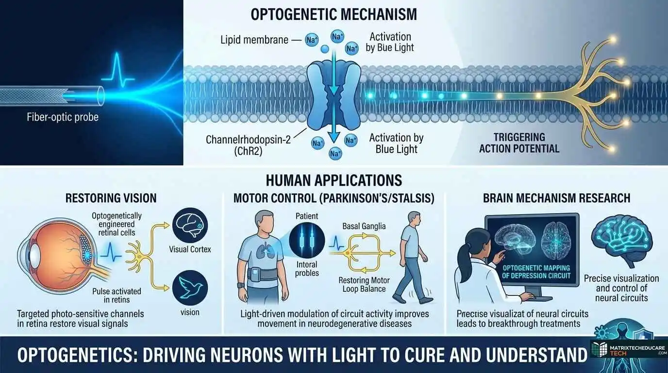
The critical opsin is channelrhodopsin-2 (ChR2), discovered by Peter Hegemann and Georg Nagel in the green alga Chlamydomonas reinhardtii . ChR2 is a light-gated cation channel: when blue light hits it, positively charged ions rush into the cell, depolarizing the membrane and — if the cell is a neuron — triggering an action potential. Karl Deisseroth and colleagues showed in 2005 that you could put the gene for ChR2 into mammalian neurons and use pulses of blue light to drive those cells to fire with millisecond precision [4]. Ed Boyden, in the same lab, demonstrated that ChR2 could be used to reliably trigger trains of action potentials in neurons .
That 2005 paper is the one most people point to as the birth of optogenetics. The term itself was coined by Deisseroth’s group in 2006 .
How It Actually Works, Step by Step
Here’s the full chain, simplified but accurate:
- Choose your opsin. Different opsins respond to different wavelengths and do different things (see the next section). You pick one based on whether you want to activate or inhibit neurons, and what color of light you’ll use.
- Deliver the gene. You package the opsin gene into a viral vector — usually an adeno-associated virus (AAV) — and deliver it to the brain region or tissue you’re studying. A genetic “promoter” sequence ensures the opsin is only expressed in the specific cell type you care about.
- Wait for expression. The cells produce the opsin protein, which inserts itself into their membranes. This takes days to weeks.
- Shine light. You implant a fiber-optic cable or use an LED and deliver pulses of light of the right wavelength. The opsin responds instantly — milliseconds — and the targeted neurons fire (or fall silent).
- Observe the effect. You record behavior, electrical activity, imaging signals, or whatever you’re measuring. Turn the light off, and the effect stops.
The elegance is in the combination: genetic specificity (only certain cells get the opsin) plus temporal precision (light turns on and off in milliseconds) plus spatial control (you aim the light where you want). None of the older methods — electrodes, drugs, lesions — combined all three.
Think of it like a light switch wired to a specific room in a vast building. Before optogenetics, you could cut power to a whole floor and see what went dark. Now you can flip the switch in one office and watch what happens to the person working there.
The Toolkit: Opsins, Light Sources, and Delivery Methods
The Opsin Family
The optogenetic toolbox has expanded enormously since ChR2. Researchers have discovered and engineered dozens of opsins, each tuned for different jobs [7]. Here’s a practical breakdown:
| Opsin | Light | Action | Best For |
| ChR2 | Blue (~470 nm) | Excitatory (cation channel) | Fast activation, standard workhorse |
| Chrimson | Red (~590 nm) | Excitatory (cation channel) | Deep tissue, dual-color experiments |
| VChR1 | Orange (~535 nm) | Excitatory (cation channel) | Independent activation alongside ChR2 |
| Halorhodopsin (NpHR) | Yellow (~570 nm) | Inhibitory (chloride pump) | Silencing neurons |
| Arch | Green (~520 nm) | Inhibitory (proton pump) | Silencing, less chloride toxicity |
| STORM | Blue | Step-function opsin | Sustained activation with brief light |
Excitatory opsins like ChR2 open channels that let positive ions in, depolarizing the cell and making it fire. Inhibitory opsins like halorhodopsin pump chloride in or protons out, hyperpolarizing the membrane and making the cell go quiet . Some opsins, called step-function opsins, stay open after a brief light pulse and can be closed with a different wavelength — useful for sustained activation without constant illumination.
A key engineering goal has been red-shifted opsins. Red light penetrates tissue more deeply than blue (think of how red light reaches your hand when you hold a flashlight through it, but blue scatters). Red-shifted opsins like Chrimson let researchers activate neurons deeper in the brain without invasive fiber optics — a major practical advantage for both research and eventual clinical use .
Light Delivery: Getting Photons to the Cells
Light doesn’t travel far through tissue on its own. In the brain, blue light penetrates only about a millimeter. To reach deeper structures, researchers use:
- Optical fibers implanted directly into the brain — the standard approach for animal research
- LEDs mounted on the skull or integrated into flexible probes
- Wireless miniaturized devices that let freely moving animals be stimulated without tethering
- Two-photon microscopy — a technique using infrared light that penetrates deeper and allows activation of individual cells with cellular resolution
For clinical applications, the delivery challenge is even greater. In the vision-restoration trials, the approach is different: the eye is accessible to external light, so patients wear special goggles that project patterned light onto the retina, activating opsin-expressing cells [9]. No fiber-optic implants needed — a critical simplification for clinical translation.
Genetic Targeting: The Specificity Engine
The real power of optogenetics isn’t just the light — it’s the genetic targeting. By choosing the right viral vector, promoter, and delivery route, you can make the opsin appear only in specific cell types — say, only parvalbumin-positive interneurons, or only dopaminergic neurons in the midbrain. This is what lets researchers ask causal questions: “What happens if I silence only the inhibitory cells in this circuit?” rather than “What happens if I silence everything in this area?”
The same logic extends to other biological systems. Optogenetics isn’t just for neurons anymore — it’s being used in heart cells, immune cells, and even cancer cells .
Applications: What Optogenetics Has Taught Us So Far
Mapping Neural Circuits
The foundational application is circuit mapping. Before optogenetics, neuroscientists knew that brain region A connected to region B, but they couldn’t easily test whether that connection was causal for a behavior. With optogenetics, you can activate a specific projection from A to B and watch the animal react — or silence it and see what stops working. This has produced a wave of causal insights into how neural circuits generate behavior, perception, and emotion.
For example, optogenetics helped identify specific circuits involved in anxiety (activating certain basolateral amygdala projections reduces anxiety-like behavior in mice), reward (dopamine neuron activation drives seeking behavior), and aggression. These aren’t correlations — they’re causal demonstrations that this specific pathway, when active or silent, produces this specific outcome.
Understanding Brain Disorders
Optogenetics has been particularly powerful for dissecting the circuit basis of neurological and psychiatric conditions. By recreating or correcting specific patterns of neural activity in animal models, researchers have gained insight into:
- Parkinson’s disease — identifying which circuits are disrupted by dopamine loss and how deep brain stimulation works at a circuit level
- Epilepsy — using optogenetics to detect and halt seizures in real time by silencing excitatory neurons or activating inhibitory ones
- Depression — mapping the circuits underlying depressive behavior and identifying rapid-acting antidepressant mechanisms
- Addiction — showing how specific projections encode drug-seeking and relapse
- Memory disorders — manipulating “engram” cells (the neurons that store specific memories) to understand how memories are formed, stored, and recalled
A striking 2026 study used optogenetics to show that reactivating specific memory “engram” cells in the retrosplenial cortex during NREM sleep accelerated memory consolidation — essentially demonstrating that you can strengthen a memory by reactivating the right neurons during the right sleep stage. This kind of causal, temporally precise experiment was simply impossible before optogenetics.
Beyond the Brain: Cardiology, Cancer, and More
While neuroscience is the dominant application, optogenetics applications have spread far beyond it:
- Cardiology — Researchers have used optogenetics to control heart rhythm with light, potentially offering a more precise alternative to electrical defibrillation
- Cancer research — Optogenetic tools are being used to interrogate signaling pathways, control gene expression, and even improve cell-based cancer therapies by providing light-regulated on/off switches for therapeutic cells.
- Cell biology — Controlling protein interactions, gene expression, and intracellular signaling with light, allowing researchers to dissect the timing and localization of cellular processes
- Plant science — Engineering light-controlled processes in plants for research and potential agricultural applications
The Clinical Frontier: Restoring Vision
The most advanced clinical application of optogenetics — and the one that moved it from the lab bench toward the bedside — is vision restoration.
The logic is elegant. In conditions like retinitis pigmentosa, the photoreceptors (the light-sensing cells of the retina) degenerate and die. But the retinal ganglion cells — the neurons that connect the eye to the brain — often survive. If you could make those surviving cells light-sensitive, you could bypass the dead photoreceptors and restore some vision.
That’s exactly what optogenetic therapy does. A viral vector delivers an opsin gene (like Chrimson) to the surviving retinal cells, making them responsive to light. The patient wears specialized goggles that convert visual scenes into specific wavelengths of light that activate the opsin .
The proof of concept came in 2021, when a single blind patient showed partial visual recovery after optogenetic therapy. Then, in October 2026, an international team led by University of Pittsburgh scientists published results in the New England Journal of Medicine showing that the therapy was safe in all 10 participants (the primary endpoint) and that six patients showed clinically meaningful increases in light sensitivity. Several improved on practical tasks — detecting objects, finding doorways, following lines. Brain recordings confirmed visual signals were reaching the cortex.
This wasn’t full vision restoration. The patients didn’t see the world the way someone with intact photoreceptors does. But it was a meaningful step — demonstrating that even in advanced retinal degeneration, some visual function can be recovered. And the approach is mutation-agnostic: because it doesn’t target a specific genetic mutation but rather makes surviving cells light-sensitive, it could potentially work for many different forms of inherited blindness .
A separate optogenetic gene therapy (sonpiretigene isteparvovec) is now heading directly to a Phase 2 trial in geographic atrophy, another form of retinal degeneration, based on promising early data.
A Quick Reference: Where Optogenetics Is Being Applied
| Field | What It’s Used For | Maturity |
| Systems neuroscience | Circuit mapping, causal behavior studies | Mature (standard tool) |
| Psychiatric disease models | Depression, anxiety, addiction circuits | Advanced research |
| Neurological disease models | Parkinson’s, epilepsy, stroke recovery | Advanced research |
| Memory research | Engram manipulation, sleep consolidation | Active frontier |
| Vision restoration | Retinitis pigmentosa, geographic atrophy | Clinical trials |
| Cardiology | Light-controlled pacing, defibrillation | Early research |
| Cancer research | Pathway interrogation, cell therapy control | Early research |
| Cell biology | Signaling, gene expression control | Growing |
The Challenges and Limitations Nobody Talks About
For all its power, optogenetics has real limitations, and anyone working with it — or investing in it — should understand them honestly.
The Light Delivery Problem
The biggest practical hurdle remains getting light to the target cells. Blue light scatters within a millimeter or two of tissue. Red light does better but still fades. For deep brain structures, you need implanted fibers, which are invasive and can damage tissue. This is why the vision applications have moved fastest — the eye is accessible to external light. For brain applications, non-invasive light delivery remains a major engineering challenge.
The Viral Vector Problem
Delivering the opsin gene requires a viral vector, and the immune system can react to viral proteins. In the brain, this is less of a concern because the brain is immune-privileged, but for repeated or systemic delivery, immune responses are a real barrier. Scaling up production of clinical-grade viral vectors is also expensive and technically demanding.
The Specificity Problem
Genetic targeting is powerful but not perfect. A promoter that’s supposed to drive expression only in one cell type may have off-target effects. The opsin may express at variable levels, making some cells more responsive than others. And in clinical applications, the concern is broader: if you make retinal ganglion cells light-sensitive, you’ve changed their fundamental biology. Long-term safety data is still being collected .
The Translation Gap
Most optogenetic research is in animals. Moving to humans introduces enormous complexity — not just technically but ethically and regulatorily. The 2026 Nature Neuroscience roadmap paper lays this out carefully: the selection of clinical indications, molecular and optical strategies for specificity, safety and regulatory navigation, and the need for careful ethical consideration are all live challenges .
Importantly, the authors note that much of optogenetics’ clinical impact will be indirect — not by shining light into human brains, but by using the causal knowledge gained from optogenetic research to develop better drugs, better stimulation targets, and better therapies. The vision trials are the direct application; the indirect applications may be broader and equally important.
The Ethical Horizon
When a technology lets you control behavior with light, ethical questions follow. In animal research, these are manageable. In humans, they become profound. Could optogenetics be used to alter behavior without consent? Could it be weaponized? Could it create unequal access to cognitive enhancement? These aren’t reasons to stop the research — they’re reasons to have the conversations alongside it, which the Nobel committee and the field’s leaders are actively encouraging .
Getting Started With Optogenetics : A Practical Guide for Researchers
If you’re a grad student, postdoc, or PI considering adding optogenetics to your work, here’s a practical roadmap drawn from the literature and the standard practices in the field.
Step 1: Define Your Question
Optogenetics is a tool, not a question. Before anything else, define what you want to know. Are you trying to identify which cells cause a behavior? Test whether a specific projection is necessary and sufficient? Map a circuit? Your question determines everything else.
Step 2: Choose Your Opsin
- Need to activate neurons? Start with ChR2 (or its faster variant, ChETA) for standard work.
- Need to silence neurons? Use halorhodopsin (eNpHR3.0) or ArchT.
- Need deep activation without invasive fibers? Consider red-shifted opsins like Chrimson.
- Need two independent populations controlled separately? Pair ChR2 (blue) with Chrimson (red) for dual-color experiments.
Step 3: Choose Your Delivery Strategy
- AAV vectors are the standard for gene delivery. Choose a serotype that targets your tissue (AAV2 for retina, AAV5 or AAV9 for broader neural expression).
- Promoter selection determines cell-type specificity. Use cell-type-specific promoters (e.g., CaMKII for excitatory neurons, GAD67 for inhibitory) or Cre-dependent systems with transgenic mouse lines for more precise targeting.
- Injection — for brain applications, stereotactic injection of the viral vector into the target region, followed by 2–4 weeks of expression time.
Step 4: Choose Your Light Delivery
- Implanted optical fibers for deep brain stimulation in freely moving animals.
- LEDs for surface or retinal applications.
- Wireless systems for experiments requiring untethered behavior.
Step 5: Validate Expression
Before running experiments, confirm that your opsin is expressed in the right cells and at sufficient levels. Use fluorescent tags (most opsins come with a fluorescent fusion protein) and histology to verify expression patterns. Test light-evoked responses with electrophysiology in a subset of animals.
Step 6: Run Controls
Controls are critical in optogenetics:
- No-virus controls — animals that went through the same surgery but didn’t get the opsin gene, to control for the effects of light itself.
- No-light controls — animals with the opsin that didn’t receive light stimulation.
- Behavior controls — ensure that the light isn’t causing heating or other non-specific effects.
Step 7: Analyze With Appropriate Statistics
Optogenetic experiments often involve within-subject comparisons (same animal, light on vs. light off). Use appropriate statistical models for repeated measures, and be transparent about multiple comparisons. The field has matured significantly in its statistical standards, and reviewers expect rigor here.
Practical Tips From the Literature
- Expression levels matter. Too little opsin and you won’t get enough current to drive firing. Too much and you can get cellular toxicity. Titrate your viral dose.
- Light power matters. Too much light can heat tissue, especially with blue wavelengths. Stay within established safe limits (typically <5 mW at the fiber tip for chronic implants).
- Off-target effects are real. Always check for opsin expression in unintended regions and account for it in your interpretation.
- Temporal precision is a feature, not a bug. Use short pulses (1–5 ms) to mimic natural firing patterns. Sustained depolarization can cause unnatural activation and cellular stress.
FAQs
- What is optogenetics?
Optogenetics is a technique that combines genetics and light to control the activity of specific cells — usually neurons — with millisecond precision. It involves introducing light-sensitive proteins (opsins) into target cells and then using light to turn them on or off .
- Who invented optogenetics?
The key figures are Peter Hegemann and Georg Nagel, who discovered channelrhodopsin in algae, and Karl Deisseroth and Ed Boyden, who demonstrated its use in mammalian neurons in 2005 The 2026 Nobel Prize was awarded for this work.
- What are opsins?
Opsins are light-sensitive proteins found in microbes and animals. In optogenetics, microbial opsins like channelrhodopsin and halorhodopsin are used to make cells responsive to specific wavelengths of light.
- What is channelrhodopsin?
Channelrhodopsin-2 (ChR2) is a light-gated ion channel from green algae. When blue light hits it, it opens and lets positive ions flow into the cell, depolarizing it and triggering an action potential in neurons .
- How does optogenetics control neurons?
Light activates the opsin protein in the cell membrane, which either opens an ion channel (causing the neuron to fire) or pumps ions (causing it to go silent), depending on the opsin used. This happens in milliseconds.
- What color light is used in optogenetics?
It depends on the opsin. ChR2 responds to blue light (~470 nm). Halorhodopsin responds to yellow light (~570 nm). Red-shifted opsins like Chrimson respond to red/orange light (~590 nm), which penetrates tissue more deeply.
- Is optogenetics used in humans?
Yes, in clinical trials. The most advanced application is vision restoration for retinitis pigmentosa. A 2026 study showed an optogenetic therapy was safe and improved visual function in some blind patients.
- . Can optogenetics cure blindness?
It’s not a cure yet, but it has shown the ability to partially restore vision. In 2026, six of ten treated patients showed meaningful improvements in light sensitivity and practical visual tasks.
- What is the difference between optogenetics and electrical stimulation?
Electrical stimulation activates all nearby cells indiscriminately. Optogenetics activates only cells that express the opsin, allowing precise, cell-type-specific control. Optogenetics also offers millisecond temporal precision that drugs can’t match.
- What animals are used in optogenetics research?
Mice and rats are the most common, but optogenetics has been used in flies, worms, fish, monkeys, and even in human tissue in clinical trials.
- Is optogenetics safe?
In animal research, it’s well established and generally safe with proper controls. In human clinical trials, the vision therapy showed a good safety profile in all 10 participants . Long-term safety data is still being collected.
- How is the opsin gene delivered to cells?
Usually through a viral vector — most commonly an adeno-associated virus (AAV) that carries the opsin gene under a cell-type-specific promoter. The virus is injected into the target tissue, and the cells produce the opsin protein over days to weeks.
- What are inhibitory opsins?
Inhibitory opsins like halorhodopsin (NpHR) and Arch pump chloride ions in or protons out of the cell, hyperpolarizing the membrane and preventing the neuron from firing. They’re used to silence specific neurons with light.
- Can optogenetics be used to control behavior?
Yes. By activating or silencing specific circuits in freely moving animals, researchers have demonstrated causal links between specific neural activity patterns and behaviors like movement, feeding, anxiety, and memory .
- What is a channelrhodopsin variant?
Researchers have engineered many ChR2 variants for improved properties — faster kinetics (ChETA), higher light sensitivity (CatCh), red-shifted activation (Chrimson), or step-function behavior (STORM) — each suited to different experimental needs.
- What are the limitations of optogenetics?
Key limitations include light delivery (light doesn’t penetrate deep tissue easily), viral vector immune responses, potential off-target expression, and the challenge of translating animal findings to humans.
- How deep can light penetrate the brain?
Blue light penetrates only about 1 mm. Red light reaches somewhat deeper. For deep brain structures, implanted optical fibers are typically required.
- Can optogenetics be used wirelessly?
Yes. Wireless miniaturized optoelectronic devices have been developed that allow stimulation of freely moving animals without tethering to fiber-optic cables.
- What is the Nobel Prize connection?
The 2026 Nobel Prize in Physiology or Medicine was awarded for the discovery of optogenetics, recognizing the contributions of Hegemann, Nagel, Deisseroth, and Boyden to developing the technique.
- How is optogenetics used in cancer research?
Optogenetic tools are used to control signaling pathways, gene expression, and protein interactions in cancer cells, allowing researchers to study how specific molecular events drive cancer progression. They’re also being explored to improve cell-based cancer therapies.
- Can optogenetics help with Parkinson’s disease?
In research, optogenetics has helped identify the circuits disrupted in Parkinson’s and how therapies like deep brain stimulation work. Direct clinical application for Parkinson’s isn’t yet available but is an active research area.
- What is an “engram” in optogenetics?
An engram is the physical trace of a memory in the brain — the specific ensemble of neurons that were active during learning. Optogenetics allows researchers to tag and reactivate these engram cells to study memory formation and recall .
- How does optogenetic vision therapy work?
A viral vector delivers an opsin gene to surviving retinal cells (like ganglion cells) in patients with photoreceptor degeneration. The patient then wears goggles that project specific wavelengths of light onto the retina, activating the opsin-expressing cells and sending visual signals to the brain [9].
- Is the vision restoration permanent?
The therapy’s effects depend on the opsin continuing to be expressed and the surviving retinal cells remaining functional. Long-term durability is still being studied, but the approach is designed to be a one-time treatment with sustained expression.
- Can optogenetics be used for psychiatric disorders?
In animal models, yes — it has provided causal insights into depression, anxiety, and addiction circuits. Direct clinical use for psychiatric disorders is further off, but the knowledge gained is informing drug development and brain stimulation therapies [12].
- What is the difference between optogenetics and chemogenetics?
Optogenetics uses light to control cells; chemogenetics uses engineered receptors activated by synthetic drugs (like DREADDs). Optogenetics offers better temporal precision; chemogenetics is less invasive since it doesn’t require implanted light sources.
- Are there any approved optogenetic therapies?
Not yet fully approved. The vision therapy is in clinical trials and heading toward Phase 2 for some indications [14]. No optogenetic therapy has received full regulatory approval as of 2026.
- What are the ethical concerns of optogenetics?
Concerns include the potential for behavioral control without consent, unequal access to cognitive enhancement technologies, and the implications of directly manipulating brain activity in humans. The field is actively discussing these issues [11].
- How expensive is optogenetic equipment?
For a research lab, the core costs include viral vectors (a few hundred to a few thousand dollars per construct), fiber-optic hardware and lasers/LEDs (several thousand dollars), and surgical equipment. It’s accessible to most well-funded academic labs.
- What’s next for optogenetics?
Key directions include red-shifted and more sensitive opsins for deeper, less invasive stimulation; improved viral vectors for clinical delivery; expansion into new clinical indications beyond vision; and indirect clinical impact through knowledge-driven drug and therapy development.
A Light Switch for the Mind
Optogenetics is one of those rare technologies that didn’t just add a new tool to the toolbox — it opened a door that was previously locked. For the first time, neuroscientists could control specific cells in living brains with the precision of a light switch, and walk through the resulting behavior to understand what those cells actually do. That capability has produced thousands of papers, rewired our understanding of neural circuits, seeded clinical trials that are already restoring partial vision to blind patients, and now earned the highest recognition in science [1][2].
But the honest story of optogenetics is also a story about patience and convergence. The opsin proteins were discovered in algae by biophysicists who had no interest in neuroscience. The viral delivery methods came from gene therapy. The fiber-optic technology came from telecommunications. The laser systems came from physics. None of these fields knew they were building toward the same thing, and it took decades for someone to put the pieces together. That’s how real breakthroughs usually happen — not in a single flash of genius, but in the slow assembly of tools from unexpected places until suddenly the impossible becomes routine.
The clinical frontier is where the next chapter gets interesting. The vision trials have proven that optogenetics can work in humans — not perfectly, not yet as a full cure, but meaningfully. The roadmap paper in Nature Neuroscience makes clear that the broader clinical impact may be indirect: the causal knowledge gained from optogenetic research feeding into better drugs, better stimulation targets, better therapies for conditions that have resisted treatment for decades [11]. That indirect path may ultimately help more people than the direct one.
If there’s a takeaway from the optogenetics story, it’s that understanding the brain requires tools precise enough to match its complexity — and that the right tools, once they exist, change everything. The next decade will see optogenetics mature from a research revolution into a clinical one, with all the challenges and promises that transition entails. The light is on. The work is just beginning.
Your call to action: If this piece helped you understand optogenetics, share it with someone who’s curious about brain science — a student, a colleague, a friend who keeps asking what that Nobel Prize was actually about. If you’re a researcher, the references throughout this post are your starting point for going deeper. And if you or someone you know is affected by inherited retinal disease, ask your ophthalmologist about ongoing optogenetic clinical trials — the science is moving, and the patient registries are open. The best way to honor a breakthrough is to use it.
⚕️Medical Disclaimer:
The information provided on this website is for general educational and informational purposes only and is not intended as a substitute for professional medical advice, diagnosis, or treatment. Always seek the advice of your physician or other qualified health provider with any questions you may have regarding a medical condition. Never disregard professional medical advice or delay in seeking it because of something you have read on this website.