

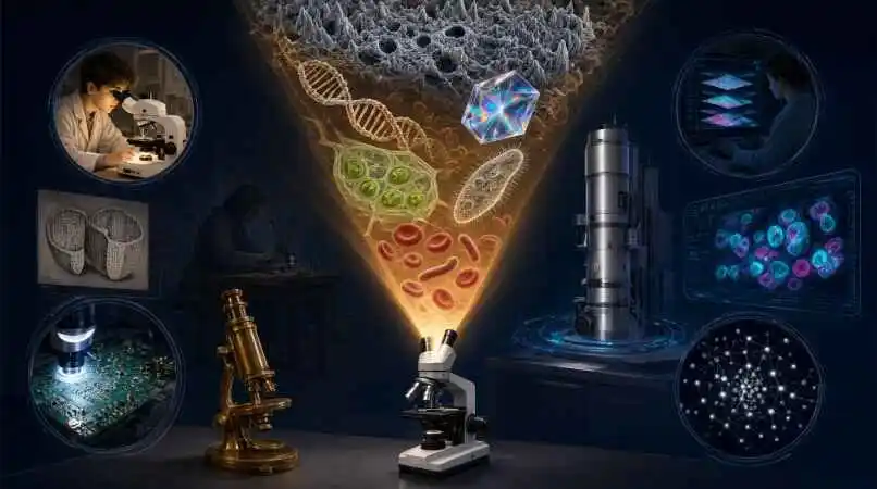
The Invisible World in Focus: A Complete Guide to Microscopes and Microscopy
Why the Smallest Things Matter Most
Every major scientific breakthrough—from the discovery of cells to the development of mRNA vaccines—has depended on our ability to see what the naked eye cannot. Microscopes are the instruments that have unlocked that invisible universe, and microscopy is the discipline that has grown around them.
Whether you are a student peering through a compound microscope for the first time, a researcher troubleshooting fluorescence signals, or a hobbyist wondering which microscope to buy, this guide covers the essentials, the nuances, and the questions people actually ask.
We will move from the history of the instrument to the technologies shaping its future, and we will finish with 30 frequently asked questions that address the practical, the technical, and the occasionally confusing aspects of microscopy.
A Brief History of Seeing the Unseen
The story of the microscope is a story of curiosity compounded by craftsmanship. The properties of concave and convex lenses were described around the year 1000, though magnifying lenses may have been used far earlier. By the 14th century, lens grinding had developed in Italy and spectacles were improving eyesight.
The pivotal moment arrived around 1590, when Dutch lens grinders Hans and Zacharias Janssen placed two lenses in a tube and created what is generally recognized as the first compound microscope. A few decades later, Robert Hooke published Micrographia in 1667, bringing microscopic observations—including the description of cork and its cellular structure—to a wide audience.
Then came Antonie van Leeuwenhoek, a Dutch tradesman whose self-made single-lens microscopes achieved magnifications of up to 270× and remained unsurpassed for over 150 years. He was the first to observe bacteria, protozoa, and human sperm, earning him recognition as the father of microbiology.
The 19th century brought mathematical rigor. Ernst Abbe formulated the relationship between resolution and the wavelength of light in 1878, making it possible to calculate the maximum resolution of a microscope. In 1932, Frits Zernike invented the phase-contrast microscope, enabling the study of transparent biological specimens without staining. Ernst Ruska followed in 1938 with the electron microscope, dramatically expanding the boundaries of resolution.
How Microscopes Actually Work: Magnification, Resolution, and Contrast
A microscope does more than make things bigger. It collects light or electrons from a specimen, manipulates that signal through lenses or detectors, and produces an image that the human eye or a camera can interpret. Three concepts govern whether that image is useful.
Magnification is the ratio of the image size to the object size. In a compound microscope, total magnification is calculated by multiplying the eyepiece magnification by the objective lens magnification. A 10× eyepiece paired with a 40× objective yields a total magnification of 400×. But magnification without resolution is empty enlargement.
Resolution is the ability to distinguish two closely spaced points as separate. It depends on the wavelength of the illumination and the numerical aperture of the objective lens. This is why electron microscopes, which use electrons with wavelengths far shorter than visible light, can resolve structures at the nanometer and even atomic scale.
Contrast is the difference in intensity between a feature and its background. Many biological specimens are nearly transparent, which is why techniques such as phase contrast, darkfield, and fluorescence microscopy exist—each is essentially a strategy for generating contrast without destroying the specimen.
A note on magnification marketing: A common misconception is that higher magnification numbers mean a better microscope. In reality, the best microscope is the one that provides the right combination of magnification, resolution, image quality, and optics for the specimen being examined. A 2000× label on a budget instrument means little if the optics cannot resolve fine detail.
The Major Types of Microscopes (and What Each One Is For)
There is no single “best” microscope. There are only microscopes suited to particular specimens, particular questions, and particular budgets. The four principal categories are optical (compound), stereo, digital/USB, and electron microscopes.
Compound (Optical) Microscopes
The compound microscope is the workhorse of laboratories, classrooms, and clinical settings. It combines an objective lens positioned close to the specimen with an ocular lens (eyepiece) to produce a magnified image. Compound microscopes are ideal for microelectronics, biotechnology, microbiology, and education. They typically offer a range of objectives—commonly 4×, 10×, 40×, and 100× oil immersion—allowing users to survey a sample at low power and then zoom in on detail.
Stereo (Dissecting) Microscopes
Stereo microscopes use two separate optical paths, producing slightly different viewing angles for each eye and generating a three-dimensional image. This makes them indispensable for dissections, surgery, botany, electronics inspection, and any task where depth perception matters. They generally operate at lower magnifications than compound microscopes but offer a much larger working distance.
Digital and USB Microscopes
Digital microscopes replace the eyepiece with a sensor and display the image on a computer screen or built-in monitor. USB microscopes are portable, plug-and-play devices that are useful for examining flat objects, printed circuit boards, and moving microorganisms. They are popular in education, quality control, and hobbyist applications where sharing images or recording video is important.
Electron Microscopes
Electron microscopes use a beam of electrons instead of light to illuminate the specimen, achieving resolutions far beyond the diffraction limit of visible light. There are two main types: scanning electron microscopy (SEM) , which detects reflected or backscattered electrons to produce detailed three-dimensional surface images, and transmission electron microscopy (TEM) , which uses electrons transmitted through an ultrathin sample to reveal internal structure at atomic resolution. SEM is widely used for surface analysis of bulk materials, while TEM excels at visualizing the internal architecture of cells and nanostructures.
Specialized Light Microscopy Techniques
Beyond the basic compound microscope, several contrast-enhancing techniques have become essential tools:
Fluorescence microscopy uses fluorophores—molecules that absorb light at one wavelength and emit it at a longer wavelength—to label specific structures with high specificity. It is the backbone of modern cell biology, enabling researchers to track proteins, visualize organelles, and quantify cellular processes.
Confocal microscopy places a pinhole in the detection path to reject out-of-focus light, enabling optical sectioning of thick specimens and three-dimensional reconstruction. It offers improved lateral resolution and is widely used in biomedical research.
Phase-contrast microscopy converts minute differences in the refractive index of transparent specimens into visible contrast, allowing live, unstained cells to be observed.
Darkfield microscopy blocks the direct illumination and collects only scattered light, making small particles and fine edges appear bright against a dark background.
Polarizing microscopy uses polarized light to detect anisotropic, birefringent structures such as crystals, fibers, and ordered molecular assemblies.
Atomic force microscopy (AFM) is a scanning probe technique that measures mechanical forces between a sharp tip and a surface, producing three-dimensional topographic maps at atomic resolution and even probing biomechanical properties of living cells.
Microscopy in the Real World: Applications Across Disciplines
Microscopy is not confined to academic research. It underpins quality control, clinical diagnosis, materials development, and education.
In biology and medicine, microscopy is used for histology, pathology, hematology, microbiology, and drug discovery. Fluorescence and confocal microscopy allow researchers to observe cellular dynamics in real time, while electron microscopy provides ultrastructural detail for tissue engineering and regenerative medicine. Correlative approaches that combine optical and atomic force microscopy are gaining traction in biomedicine, particularly for studying pathogens and cellular mechanics.
In materials science and engineering, SEM and TEM are essential for characterizing nanostructures, thin films, and composite materials. Electron microscopy supports failure analysis and quality control in industries ranging from semiconductors to aerospace.
In education, microscopes are a gateway to STEM engagement. Even simple stereo and compound microscopes allow students to observe plant cells, microorganisms, and mineral structures, building intuition for scale and biological organization.
In industry and forensics, digital microscopes and stereo microscopes are used for component inspection, trace evidence analysis, and documentation. USB microscopes have made high-resolution imaging accessible in field settings and small workshops.
How to Choose a Microscope: A Practical Buying Guide
Choosing a microscope can feel overwhelming because the specifications are numerous and the marketing claims are loud. The most important factor is your application: what do you want to look at? A microscope for observing living cells has different requirements from one for inspecting circuit boards or identifying minerals.
Magnification and resolution. Resist the temptation to buy the highest magnification number. Instead, look for quality optics and a numerical aperture that matches your intended use. For most educational and routine laboratory work, a compound microscope with 4×, 10×, 40×, and 100× objectives covers a wide range of specimens.
Eyepiece configuration. Monocular microscopes are the least expensive, followed by binocular and trinocular models. Binocular eyepieces are far more comfortable for extended use and are generally worth the investment for anyone who will spend significant time at the instrument. Trinocular models add a camera port for imaging.
Illumination. LED illumination is now standard and offers consistent color temperature, low heat, and long life. Adjustable brightness and a properly aligned condenser are important for contrast control.
Stage and focus controls. A mechanical stage allows precise, smooth movement of the specimen, which is especially important at higher magnifications. Coaxial coarse and fine focus knobs make focusing intuitive.
Digital capability. If you need to document, share, or measure images, consider a digital or trinocular microscope. USB microscopes offer plug-and-play convenience, while dedicated camera systems provide higher image quality and software integration.
Budget and future needs. Entry-level student microscopes are affordable and adequate for basic observation. Research-grade instruments cost significantly more but offer superior optics, stability, and modularity. It is often better to buy a well-made instrument with fewer features than a feature-heavy instrument with poor optics.
Keeping Your Microscope in Top Condition: Maintenance Essentials
A clean microscope produces better images, lasts longer, and reduces downtime. The fundamentals of microscope care are straightforward but frequently overlooked.
Cleaning optics. Use an air blower to remove loose dust before touching any lens. Clean eyepieces and objectives with soft lens paper and a suitable lens cleaning fluid, such as isopropanol or a commercial optical cleaning solution. Never use harsh chemicals or dry paper on optical surfaces. If immersion oil is used, wipe the objective immediately after use—dried oil residues are difficult to remove and can reduce objective lifespan.
Protecting against environmental damage. Microscopes do not tolerate humidity well. Fungal growth can damage lens coatings, and moisture can corrode mechanical components. Cover the microscope when not in use, and store it in a clean, dry, dust-free environment. In humid climates, a dehumidifier or desiccant can help protect optics.
Handling and transport. Carry a microscope with both hands—one under the base and one on the arm—and avoid tilting it more than necessary. When transporting, remove loose accessories and secure the stage.
Routine inspection. Check the condenser front lens, stage, and specimen holder for debris. Rotate objectives and move the specimen while observing to determine whether a spot is on the optics, the slide, or the camera sensor.
The Future of Microscopy: Super-Resolution, AI, and Computational Imaging
The resolution barrier that Ernst Abbe described in the 19th century is no longer absolute. Super-resolution microscopy techniques—including STED, PALM, STORM, and structured illumination—have enabled fluorescence imaging beyond the diffraction limit, and the field continues to evolve rapidly.
Recent advances combine deep learning with computational reconstruction. A 2026 study demonstrated a super-resolution system that uses precision motion modulation and a generative adversarial network (GAN) to recover sub-pixel information without modifying the existing optical system. Other work has achieved sub-60 nm spatial resolution in coherent Raman scattering microscopy using adaptive pointillist deconvolution, a purely computational advance.
Single-frame computational super-resolution methods are also maturing. Researchers have reconstructed super-resolved images directly from a single diffraction-limited camera frame using deep learning, resolving emitters separated by 35 nm at a wavelength of 580 nm—a sevenfold improvement beyond the Rayleigh criterion.
These developments are significant because they reduce the hardware burden of super-resolution imaging. Instead of requiring specialized optics or complex sample preparation, computational approaches can extract more information from conventional microscopes, bringing nanoscale imaging within reach of a wider range of laboratories.
At the same time, AI is improving image denoising, segmentation, and analysis, making it easier to extract quantitative data from microscopy images and reduce the subjectivity of visual interpretation.
FAQs
- What is the difference between a microscope and microscopy?
A microscope is the instrument; microscopy is the field of study and practice that uses microscopes to observe and analyze specimens. - What magnification do I need for looking at cells?
Most animal and plant cells are visible at 100× to 400× total magnification, though finer details require 400× to 1000× with good optics and proper staining. - Why do I need to start with the lowest magnification objective?
The 4× objective provides the widest field of view, making it easier to locate the specimen and bring it into rough focus before switching to higher powers. - How do I calculate total magnification?
Multiply the eyepiece magnification by the objective lens magnification. A 10× eyepiece with a 40× objective gives 400× total magnification. - What is the difference between magnification and resolution?
Magnification enlarges the image; resolution is the ability to distinguish fine detail. High magnification without good resolution produces a large but blurry image. - What is numerical aperture (NA)?
NA is a measure of the light-gathering ability of an objective lens. Higher NA generally means better resolution and brighter images. - Why do some objectives use immersion oil?
Immersion oil has a refractive index similar to glass, reducing light loss between the objective and the specimen and improving resolution at high magnifications. - Can I use a compound microscope to look at solid objects?
Compound microscopes are designed for thin, translucent specimens mounted on slides. Solid objects are better viewed with a stereo microscope. - What is the difference between SEM and TEM?
SEM detects reflected or backscattered electrons to image surface topography, while TEM uses transmitted electrons through an ultrathin sample to reveal internal structure at higher resolution. - Do electron microscopes require special sample preparation?
Yes. Samples must be dehydrated, fixed, and often coated or sectioned, depending on whether SEM or TEM is used. Live specimens cannot be imaged in a standard electron microscope. - What is fluorescence microscopy used for?
It is used to visualize specific molecules, structures, or processes by tagging them with fluorescent probes, enabling high-specificity imaging in cell biology and medicine. - What is the advantage of confocal microscopy?
Confocal microscopy rejects out-of-focus light, enabling optical sectioning and three-dimensional reconstruction of thick specimens with improved contrast. - Can I view live cells with a microscope?
Yes, but the technique matters. Phase-contrast and darkfield microscopy allow observation of live, unstained cells, while fluorescence and electron microscopy typically require fixation or labeling. - How do I clean microscope lenses?
Use an air blower first, then lens paper and a suitable lens cleaning fluid. Avoid dry paper, harsh chemicals, and touching optical surfaces with bare fingers. - How often should I clean my microscope?
Clean optics after each use if immersion oil is used; otherwise, inspect and clean as needed. Cover the microscope when not in use to prevent dust accumulation. - Where should I store my microscope?
In a clean, dry, dust-free location with stable temperature and moderate humidity. A dust cover is essential. - What is the purpose of the condenser?
The condenser focuses light onto the specimen and controls the cone of illumination, which affects contrast and resolution. - What is Köhler illumination?
Köhler illumination is an alignment procedure that ensures even, focused illumination of the specimen and minimizes stray light, producing optimal contrast and resolution. - Why does my image look blurry even at high magnification?
Possible causes include dirty optics, incorrect focus, insufficient illumination, a thick specimen, or an objective with low numerical aperture. Resolution, not magnification alone, determines sharpness. - What is a stereo microscope used for?
Stereo microscopes provide three-dimensional views of solid objects and are used for dissection, electronics inspection, botany, and any task requiring depth perception. - Can I attach a camera to my microscope?
Yes. Trinocular microscopes have a dedicated camera port. Monocular and binocular microscopes can often accept cameras with adapters, though the setup may be less convenient. - What is the difference between brightfield and darkfield microscopy?
Brightfield microscopy illuminates the specimen directly and shows dark features against a bright background. Darkfield microscopy blocks direct light and collects only scattered light, making features appear bright against a dark background. - What is phase-contrast microscopy best for?
It is ideal for observing transparent, unstained specimens—such as living cells in culture—by converting refractive index differences into visible contrast. - What is polarizing microscopy used for?
It is used to identify birefringent materials, including crystals, minerals, fibers, and ordered biological structures. - What is atomic force microscopy (AFM)?
AFM is a scanning probe technique that measures forces between a sharp tip and a surface to produce three-dimensional topographic images at high resolution. - Is super-resolution microscopy the same as electron microscopy?
No. Super-resolution microscopy uses light and computational or optical tricks to surpass the diffraction limit, while electron microscopy uses electrons and requires different sample preparation. - What is the role of AI in microscopy?
AI is used for image denoising, super-resolution reconstruction, segmentation, and automated analysis, helping researchers extract more information from images and reduce manual effort. - How do I measure a specimen with a microscope?
Use an eyepiece reticle calibrated against a stage micrometer. The reticle divisions are converted to real measurements for each objective-eyepiece combination. - What are the most common mistakes beginners make?
Starting at high magnification, using dirty slides, forcing focus knobs, neglecting illumination adjustment, and failing to cover the microscope when not in use. - Do I need an expensive microscope to get started?
No. A well-made entry-level compound or stereo microscope with good optics and comfortable eyepieces is sufficient for most educational and hobbyist applications. Prioritize optical quality and build quality over inflated magnification claims.
A Tool That Rewards Curiosity
The microscope is one of the few instruments that has fundamentally changed how humans understand life, matter, and disease. From Leeuwenhoek’s single-lens marvels to today’s AI-assisted super-resolution systems, the pursuit of seeing smaller and clearer has never stopped.
Whether you are choosing your first microscope or refining a research workflow, the principles remain the same: match the instrument to the question, prioritize resolution and contrast over magnification numbers, and take care of your optics. The invisible world is patient. It has been waiting for centuries, and it will reward the curiosity you bring to it
Disclaimer:
The information provided in this article is for general educational and informational purposes only. It is not intended as medical advice, diagnosis, or treatment, and it should not be used as a substitute for consultation with a qualified healthcare professional.